
What does a "thickened endometrium" actually mean?
The endometrium is the inner lining of the uterus. "Thickened" describes what the ultrasound measured. It can be caused by hormonal effects, polyps, endometrial hyperplasia or, less often, cancer, and ultrasound cannot reliably tell these apart. Only examining a tissue sample can.[2]
Before menopause the lining thickens and sheds with every cycle, so its thickness depends on the day of the cycle. After menopause, oestrogen levels fall and the lining is normally thin. A thicker lining after menopause has several possible explanations:
- Hormonal effect: oestrogen therapy or medicines such as tamoxifen can stimulate the lining.
- Endometrial polyps: benign growths from the lining, which can also cause spotting.
- Endometrial hyperplasia: an abnormal build-up of lining cells, some types of which are precancerous.
- Endometrial cancer: one of the possibilities that testing is meant to confirm or exclude.
Ultrasound also has limits. Obesity, fibroids, adenomyosis, a tilted uterus or earlier uterine surgery can make the lining hard to measure reliably.[2] That is why the report should say whether the endometrium was clearly and fully seen.
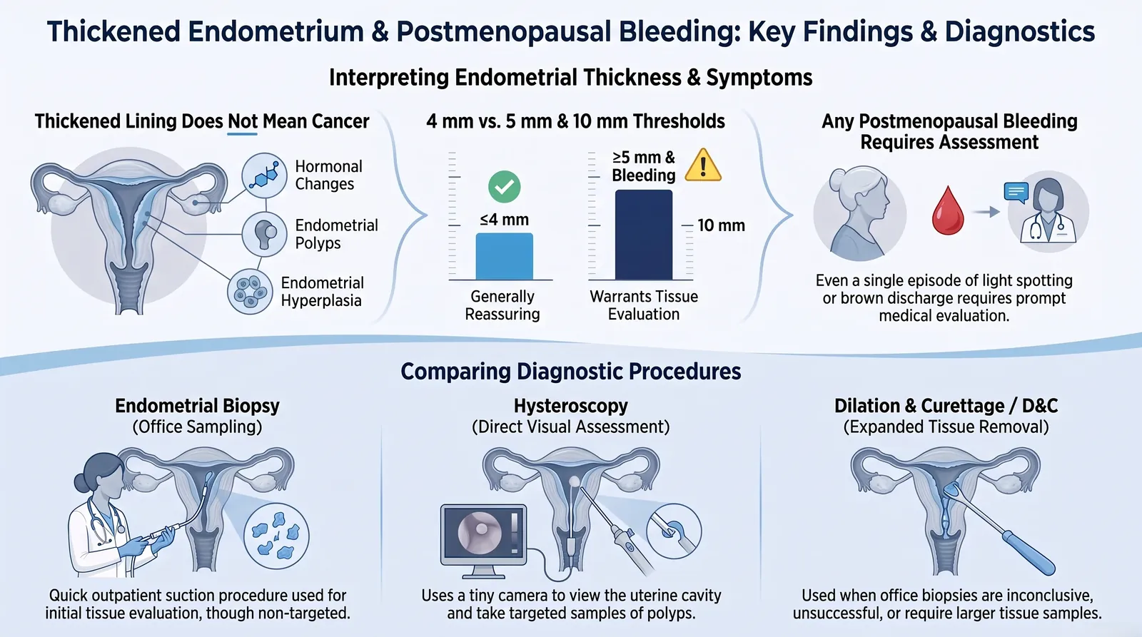
The infographic labels are in English. Note: since April 2026 ACOG advises tissue sampling together with ultrasound for most women with postmenopausal bleeding, even when the lining is 4 mm or less.
An ultrasound report that says "thickened endometrium", "endometrial thickness 5 mm" or "10 mm" is frightening to read, especially after bleeding in the years after menopause. The honest answer is that a thick lining is a finding that needs interpreting, not a cancer diagnosis. This guide explains what the numbers mean, what the symptoms and risk factors of uterine (endometrial) cancer are, and how endometrial biopsy, hysteroscopy and D&C differ. Unfamiliar terms are explained in the A to Z glossary.
Any bleeding after menopause, even a single spot, needs a doctor's assessment. If bleeding comes back, it needs fresh evaluation even when an earlier ultrasound showed a thin lining.
What do 4 mm, 5 mm and 10 mm endometrial thickness mean?
There is no single "normal" number for every woman. What matters most is whether you have had bleeding after menopause. With bleeding, a clearly seen lining of 4 mm or less has traditionally been reassuring, and anything above 4 mm needs further evaluation. Without bleeding, a thick lining found by chance is assessed case by case, and a measurement above 4 mm does not automatically need a biopsy.[2]
| Situation | What it generally means |
|---|---|
| After menopause, with bleeding, 4 mm or less (fully seen) | Cancer is unlikely, but not impossible. Recurrent or persistent bleeding still needs tissue testing. |
| After menopause, with bleeding, 5 mm | Above the commonly used 4 mm threshold. Further evaluation is needed, usually endometrial sampling. It is not a cancer diagnosis. |
| After menopause, with bleeding, 10 mm | Needs prompt tissue testing, and sometimes hysteroscopy. Most women with postmenopausal bleeding do not have cancer, but this finding must be properly investigated. |
| After menopause, no bleeding, more than 4 mm found by chance | Does not routinely trigger a biopsy. Your risk factors and how the lining looks on ultrasound guide the decision.[2] |
| After menopause, no bleeding, about 10 to 11 mm or more | Some studies use about 11 mm as a point where further investigation is reasonable. It is not a universal rule, and the decision is individual.[7] |
| Before menopause | Thickness changes through the cycle, so 5 mm or 10 mm can be normal. It is read together with the cycle day, symptoms and age. |
What changed in 2026?
Earlier guidance allowed ultrasound alone when the lining measured 4 mm or less. In April 2026 the American College of Obstetricians and Gynecologists (ACOG) updated this: most women with postmenopausal bleeding should have both a transvaginal ultrasound and endometrial tissue sampling at the first evaluation. ACOG noted that recent data suggest ultrasound-only evaluation may miss around 5% to 12% of cancers.[1]
Biopsy may be left out only when all of these are true: a single episode of bleeding; an endometrium fully seen and measuring 4 mm or less; no strong risk factors for endometrial cancer; the woman has been told that any further bleeding needs immediate re-evaluation; and there is no barrier to getting prompt gynaecological review.[1] Guidance differs between countries, so your own doctor's advice comes first.
"A thin lining lowers the chance of cancer. It does not remove it, and it never makes repeated bleeding safe to ignore."
Is postmenopausal bleeding always cancer, and how much bleeding is abnormal?
No, postmenopausal bleeding is not always cancer. Cancer is found in a minority of women with it, roughly 1% to 14% depending on age and risk factors.[2] But about 9 in 10 women with endometrial cancer have bleeding after menopause,[1] and no amount of bleeding is too small to check. Spotting counts.
Menopause is generally defined as 12 months without a period. Any vaginal bleeding after that point is postmenopausal bleeding, whether it is a few spots, a pink or brown stain, a light period-like bleed or a heavy flow. The most common cause is thinning (atrophy) of the lining of the vagina or uterus.[2] Other causes include endometrial or cervical polyps, infection or inflammation, the effect of hormone therapy, endometrial hyperplasia and cancer.
The amount of blood does not tell you the cause. A single light episode can come from a serious cause and heavy bleeding can come from a harmless one, which is why the aim is to find the source rather than guess. Do not wait for a second episode before seeing a doctor.
What are the symptoms of uterine and endometrial cancer?
Abnormal vaginal bleeding is the most common symptom. This can be bleeding after menopause, bleeding between periods or a change in periods. Vaginal spotting and unusual discharge, even without visible blood, can also be signs. Pelvic pain or fullness and unexplained weight loss are more often seen when the disease is more advanced.[3]
"Uterine cancer" most often means endometrial cancer, which starts in the lining. Rarer cancers such as uterine sarcoma start in the muscle wall and are different diseases. The symptoms to watch for are:
- Any bleeding or spotting after menopause
- A change in your periods, or bleeding between periods
- Watery, unusual or blood-stained vaginal discharge
- Pelvic pain, pressure or a feeling of fullness (more often later in the illness)
- Weight loss without trying (more often later in the illness)
None of these symptoms proves cancer, and each has non-cancer causes. They are reasons to be examined, not reasons to panic. For how fibroid and cancer symptoms differ, see our uterine cancer awareness guide and Fibroid or Uterine Cancer? Know the Facts, and for other warning signs, Do I Have Cancer?. The questions below answer how each symptom is judged in different situations.
Who has a higher risk of endometrial cancer?
Risk rises with exposure of the lining to oestrogen that is not balanced by progesterone. Obesity, type 2 diabetes, polycystic ovary syndrome (PCOS), tamoxifen, oestrogen taken alone without a progestogen, older age and inherited conditions such as Lynch syndrome all increase risk. Having a risk factor does not mean you will get cancer, and many women with endometrial cancer have none.[4]
- Obesity and metabolic syndrome: body fat produces oestrogen, which stimulates the lining.
- Type 2 diabetes: an established risk factor, often overlapping with obesity.
- PCOS: infrequent ovulation means the lining can be exposed to oestrogen without regular shedding.
- Tamoxifen: used for breast cancer, it can act like oestrogen on the uterus. Do not stop it without speaking to your treating doctor, and report any bleeding promptly.
- Lynch syndrome or a family history of endometrial or colorectal cancer.
- A previous diagnosis of endometrial hyperplasia.
When a woman with postmenopausal bleeding has higher-risk features, endometrial sampling is the preferred first test.[2]
Is endometrial hyperplasia cancer? Can it become cancer?
Endometrial hyperplasia is not cancer, but some types can progress to cancer or exist alongside a cancer that has not yet been found. What matters most is whether the cells are atypical (abnormal-looking). The diagnosis is made from tissue, never from thickness alone.
- Hyperplasia without atypia: has a low risk of becoming cancer. It is usually managed with progestogen treatment, attention to risk factors such as weight, and repeat biopsy to confirm it has settled.
- Atypical hyperplasia (also called endometrial intraepithelial neoplasia, or EIN): is precancerous. Cancer is often found alongside it. In pooled studies, about one in three women with atypical hyperplasia had cancer found within three months of diagnosis, and the risk of later progression is high.[5] Hysterectomy is the definitive treatment. Women who wish to preserve fertility, or who cannot have surgery, may be offered hormonal treatment with close specialist follow-up. Read the section on EIN for details.
What is EIN (endometrial intraepithelial neoplasia), and is it cancer?
EIN is a precancerous change in the lining of the uterus. It is not cancer. However, it can progress to cancer over time, and cancer may already be present somewhere in the lining even when the biopsy shows only EIN. That is why EIN needs specialist assessment and a clear treatment plan.
In EIN, some of the glands in the lining have become crowded, and their cells look abnormal (atypical) under the microscope. The word neoplasia sounds frightening, but here it means abnormal growth of cells. It does not automatically mean cancer.
Reports may use two names for the same condition: EIN or atypical endometrial hyperplasia (AEH). The name depends on the classification system the laboratory follows. The American College of Obstetricians and Gynecologists (ACOG) does not prefer one name over the other.[11]
How is EIN different from hyperplasia without atypia?
Not all endometrial hyperplasia carries the same risk. The most important question is whether the cells are atypical.
| Feature | Hyperplasia without atypia | EIN (atypical hyperplasia) |
|---|---|---|
| What the cells look like | A thicker lining with more glands, but the cells look normal | Crowded glands with abnormal-looking cells |
| Chance of becoming cancer over time | Less than 5 in 100 women over 20 years. Many cases return to normal on their own.[12] | Much higher. In one study of women with atypical hyperplasia who were treated with hormones, about 8 in 100 developed cancer within 4 years, and 27.5 in 100 by 19 years.[11] |
| Cancer found at the same time | Not measured in the pooled studies analysed; none reported it.[5] | Common enough that cancer must be ruled out before any conservative treatment (see below) |
| Usual treatment | Progestin tablets or a hormonal coil, with repeat checks | Removal of the uterus (hysterectomy) is the definitive treatment. Hormone treatment is for selected women, with close follow-up. |
How often is cancer found when EIN is diagnosed?
This is one of the most important questions for women and their families. Among women diagnosed with EIN who have their uterus removed, endometrial cancer is found in the removed uterus in approximately 30% to nearly 50% of cases.[11] Pooled studies of atypical hyperplasia also report that about one in three women had cancer found at the same time.[5]
This figure does not mean that 30 to 50 in 100 women with EIN will develop cancer in the future. It means that cancer was already present but was not seen in the original biopsy. A small biopsy samples only part of the lining, and a small area of cancer can be missed. The real chance for an individual woman depends on her biopsy, scans and examination.
For this reason, ACOG advises doctors to try to rule out cancer before choosing any conservative treatment. The most accurate way to do this is hysteroscopy, in which a thin telescope is used to look inside the uterus, with further sampling of any suspicious area.[11] A scan alone cannot rule out cancer, because only tissue examined under a microscope can answer that question.
"EIN is not cancer, but a cancer may already be hidden in the lining. Find that out before choosing a treatment."
What are the treatment options for EIN?
Treatment depends on your age, whether you have reached menopause, your general health and fitness for surgery, whether you still want children, and whether cancer has been ruled out.[11]
1. Hysterectomy: the definitive treatment
For most women with EIN, removing the uterus is the definitive treatment and is commonly recommended, particularly after menopause.[11] It removes the abnormal lining, allows the whole uterus to be examined for hidden cancer, and removes the risk of EIN progressing in that uterus. If the examination of the removed uterus shows no cancer, routine follow-up biopsies are not needed.[11]
- Total hysterectomy removes the uterus and cervix. A supracervical hysterectomy, which keeps the cervix, is not advised for EIN, because the lower end of the lesion cannot be fully checked.[11]
- Removing the ovaries is a shared decision. In women who have not yet reached menopause, the decision weighs the risks of early surgical menopause against the chance of hidden cancer. Discuss this with your surgeon.[11]
- The removed uterus should be carefully examined by a pathologist, because cancer may be hidden in it. If cancer is found, further treatment depends on its type, grade, extent and stage.
- Endometrial ablation is not advised for EIN. This procedure destroys the lining without removing the uterus, and it has high rates of persistence and recurrence for EIN.[11]
2. Progestin treatment: for selected women
Not every woman can or wants to have surgery. For women who cannot have a hysterectomy, or who want to keep their fertility, progestin treatment may be offered. It must be followed by close specialist follow-up.[11] Progestin opposes the effect of oestrogen on the lining and encourages the abnormal cells to settle. The main options are:
- Progestin tablets, taken continuously every day. Continuous treatment works better than cyclical use.[11]
- A levonorgestrel intrauterine system (LNG-IUS), a hormonal coil placed inside the uterus. Studies suggest it may lead to more regression than tablets alone, although the evidence is limited and not fully consistent.[11]
- A combination of the coil and tablets, in selected cases.[11]
Body weight and blood sugar also matter. In one study of women with obesity, those who lost more than 10% of their body weight during LNG-IUS treatment were nearly four times more likely to respond to it. Weight loss and good blood sugar control may also lower the risk of EIN and endometrial cancer.[11]
Some women who have not finished their families want to conceive. In that case, progestin treatment followed by fertility treatment may be discussed, ideally with a fertility specialist. Conditions such as PCOS, obesity and diabetes can make pregnancy harder, so these should be assessed early.[11] Our article on fertility after gynaecological cancer explains the options.
Hormone treatment does not replace follow-up. It is only appropriate after cancer has been ruled out, and only if you can return for repeat biopsies on time. If travel or other problems make regular follow-up difficult, tell your doctor before treatment starts.
How is EIN followed up after hormone treatment?
The first repeat biopsy is needed within 3 to 6 months of starting progestin treatment. It shows whether the abnormal cells are responding.[11]
The result usually falls into one of four groups:
- Complete response: EIN or atypical hyperplasia is no longer seen.
- Regression: the cells have improved to hyperplasia without atypia, but the change is not fully resolved.
- Persistence: EIN is still present with no change.
- Progression: cancer is found. This needs urgent review and usually surgery.
If there is little or no response in the first 3 to 6 months, your doctor may consider a further 3 to 6 months of treatment after discussion with you. If there is no response after 9 to 12 months in total, other options, including surgery, should be discussed.[11]
Even after a good response, EIN can come back. ACOG considers it reasonable to repeat the biopsy every 3 to 6 months for up to 2 years, and a review of studies found recurrence in about 23% of women treated for EIN.[11] This is why any bleeding during or after treatment must be reported promptly, even if the last biopsy was normal.
What does the biopsy report wording mean?
| Report wording | What it generally means |
|---|---|
| EIN, or atypical endometrial hyperplasia (AEH) | A premalignant change. It needs specialist review and a clear treatment plan. |
| Hyperplasia without atypia | Thickening of the lining with normal-looking cells. This is usually a lower-risk finding. |
| Insufficient or inadequate tissue | The sample was too small to give an answer. This is not a normal result. Ask about hysteroscopy and further sampling. |
| No evidence of malignancy | No cancer was seen in the sample. Cancer elsewhere in the lining can still be missed if the sample was limited. |
| Suspicious for EIN | Some features are concerning but not conclusive. Further sampling or a second opinion from a pathologist may be needed. |
Questions to ask your gynaecologist
- Has cancer been ruled out? Do I need hysteroscopy with further sampling?
- Given my age, health and wish for children, is hysterectomy the safest option for me?
- If I choose hormone treatment, when is my next biopsy, and who will follow me up?
- What should I do if bleeding returns during or after treatment?
Which tests diagnose uterine cancer? Is ultrasound enough?
Ultrasound is not enough on its own. It measures the lining and can show polyps or irregular areas, but it cannot confirm or exclude cancer. The diagnosis is made by examining tissue from the endometrium, usually obtained by endometrial biopsy, and by hysteroscopy with or without D&C when needed. Transvaginal ultrasound is also not a screening test for endometrial cancer in women without symptoms.[2]
1. History and examination
Your doctor asks about bleeding, menopause, hormone treatment, tamoxifen and relevant conditions, and examines you to find out whether the bleeding comes from the vagina, cervix or uterus.
2. Transvaginal ultrasound
This measures the lining and looks for polyps, fibroids or an irregular appearance. It is a triage tool, not a final answer.
3. Endometrial biopsy
A small sample of the lining is examined under the microscope. This is the test that can show hyperplasia or cancer.
4. Hysteroscopy, with or without D&C
Used when a polyp or focal lesion is suspected, when the biopsy sample is inadequate, or when bleeding continues after a negative biopsy.
5. Further tests only if cancer is found
MRI, CT and sometimes other scans are used to plan treatment. They are covered below. A Pap smear tests the cervix and cannot diagnose or exclude endometrial cancer.
Endometrial biopsy vs hysteroscopy vs D&C: what is the difference?
Endometrial biopsy takes a small blind sample using a thin suction tube and is usually the first test, done in the outpatient clinic. Hysteroscopy uses a thin telescope to look inside the uterus and take a targeted sample. D&C scrapes tissue from the cavity, usually under anaesthesia as a day-care procedure. They are steps in a sequence rather than competitors, and the next step depends on what the earlier one shows.
| Procedure | What it involves | Typically used when | Main limitation |
|---|---|---|---|
| Endometrial biopsy (for example Pipelle) | A thin flexible tube removes a small sample, usually in the clinic without general anaesthesia | First test for postmenopausal bleeding, abnormal bleeding or a suspicious ultrasound | It is a blind sample. The tissue can be insufficient, and a small focal area such as a polyp can be missed |
| Hysteroscopy | A thin telescope passes through the cervix so the cavity can be seen and sampled or a polyp removed | Suspected polyp or focal lesion, inadequate biopsy, or continuing bleeding after a negative biopsy | May cause more discomfort than a biopsy, and sometimes needs anaesthesia |
| D&C (dilatation and curettage) | The cervix is gently widened and tissue is scraped from the cavity, usually under anaesthesia | Office biopsy not possible, inadequate or inconclusive; persistent bleeding | Done without sight it can miss a focal lesion, so it is best combined with hysteroscopy |
In a large pooled analysis, the Pipelle device detected 99.6% of endometrial cancers in postmenopausal women.[6] Even so, an insufficient or failed sample is common, and ACOG recommends hysteroscopy with D&C when blind sampling is negative but bleeding persists or recurs.[2]
Is endometrial biopsy painful? Is hysteroscopy painful?
Experiences vary. A biopsy usually causes brief period-like cramps, and some women find it more uncomfortable. Hysteroscopy done in the clinic can also range from mild to quite crampy, while D&C and operative hysteroscopy are generally done under anaesthesia or sedation. Ask your doctor in advance about pain relief, and mention if an earlier attempt was very painful or unsuccessful.
Can a biopsy or hysteroscopy spread cancer?
Fear of spread should not delay a needed test. Endometrial biopsy is the standard way to reach a diagnosis, and the harm from delayed diagnosis is far greater than any theoretical concern. For hysteroscopy, some studies found more cancer cells in pelvic fluid afterwards, but recent combined analyses have found no meaningful effect on recurrence or survival.[8] Keeping the fluid pressure low during the procedure is also recommended.
Can a biopsy miss cancer? What if it is negative but bleeding continues?
Yes, no test is perfect. A biopsy can miss a small or focal abnormality, and a report of "insufficient tissue" is not a normal result. If a biopsy is negative or insufficient and bleeding continues or returns, you need further evaluation, often hysteroscopy with directed sampling or D&C, whatever the ultrasound thickness was.[2]
Some aggressive cancers, particularly the non-endometrioid types, can occur with a thin lining.[2] This is why repeated or ongoing bleeding should always be followed up, not explained away by a normal-looking scan.
Do I need an MRI, CT or PET-CT?
Not for a thickened endometrium alone. These scans are used after cancer has been diagnosed, to plan treatment. They do not replace biopsy, because the diagnosis comes from tissue.
- MRI: shows how deeply a cancer has grown into the uterine muscle and whether it has reached the cervix.
- CT scan and chest imaging: check whether the cancer has spread to lymph nodes or other organs.[4]
- PET-CT: used only in selected situations, not routinely for everyone.
What do grade 1, 2 and 3 and the different cancer types mean?
Grade describes how abnormal the tumour looks. For the most common type, endometrioid carcinoma, it depends on how much of the tumour grows in solid sheets instead of forming gland-like structures. Grades 1 and 2 are called low-grade and grade 3 is high-grade. Grade is different from stage, which describes how far the cancer has spread.[10]
| Grade | Solid growth in the tumour | Group |
|---|---|---|
| Grade 1 | 5% or less | Low-grade |
| Grade 2 | 6% to 50% | Low-grade |
| Grade 3 | More than 50% | High-grade |
- Endometrioid carcinoma: the most common type. It can be low-grade or high-grade.
- Serous carcinoma: a less common type that is considered high-grade by definition.
- Clear-cell carcinoma: a less common type, also considered high-grade by definition.
- Carcinosarcoma: a high-grade tumour with both carcinoma and sarcoma-like components, classified with the endometrial carcinomas.
Many centres now also test the tumour for molecular features (POLE mutation, mismatch-repair status, p53 status), which refine the risk group and guide treatment. The 2023 FIGO staging system includes this molecular information in early-stage disease.[10] Ask your doctor to explain your own report (our guide to understanding a cancer diagnosis can help you prepare, and our uterine biopsy report explainer covers grade, type and molecular results in more detail), because grade, type, molecular result and stage are read together.
What should I do if my report says "thickened endometrium"?
Do not ignore it and do not panic. Have the report reviewed by a gynaecologist, in person and promptly if you have had any bleeding after menopause. The next step depends on your age, menopausal status, symptoms and risk factors, not on the number alone.
Bring these to your appointment:
- The full ultrasound report, including whether the lining was fully seen and whether polyps or irregular areas were described
- Details of any bleeding, spotting or discharge, with dates
- When your last period was, or when you reached menopause
- Your medicines, especially hormone therapy or tamoxifen
- Conditions such as diabetes or PCOS, and any earlier biopsy or hysteroscopy reports
Our free symptom diary and questions-to-ask sheet can help you prepare. Useful questions to ask: Was the endometrium clearly seen? Do I need a biopsy now? What should I do if bleeding returns? If a biopsy shows atypical hyperplasia or cancer, treatment planning is best done with a gynaecological oncologist before any operation. See which doctor to see and our page on uterine cancer treatment in Kolkata.
Frequently Asked Questions
Does a thick endometrium mean cancer?
No. A thickened lining can result from hormonal effects, polyps, hyperplasia or cancer, and ultrasound cannot separate them. Tissue testing is what answers the question, and the need for it depends on whether you have bleeding, your age and your risk factors.[2]
What endometrial thickness is normal after menopause?
After menopause the lining is usually thin. For a woman with bleeding, 4 mm or less, clearly seen, is the traditional reassuring measurement. For a woman without bleeding there is no single normal cut-off, and a measurement above 4 mm found by chance does not automatically need a biopsy.[2]
What endometrial thickness is suspicious?
With postmenopausal bleeding, anything above 4 mm needs further evaluation, and ACOG's 2026 update advises tissue sampling for most women with bleeding whatever the measurement.[1] Without bleeding, about 11 mm is used in some studies as a point for further investigation, but the decision is individual.[7]
What does a 5 mm endometrium mean?
After menopause with bleeding, 5 mm is above the 4 mm threshold and needs further evaluation, usually endometrial sampling. It does not mean cancer. Before menopause, 5 mm can be a normal measurement for some days of the cycle.
What does a 10 mm endometrium mean?
With bleeding after menopause, 10 mm needs prompt tissue testing. Without bleeding, it needs an individual decision based on your risk factors and how the lining looks. It is not a universal cancer cut-off, and before menopause it can be normal.
Does a thick endometrium require a biopsy?
With postmenopausal bleeding, most women should have endometrial sampling together with ultrasound. Without bleeding, a lining above 4 mm found by chance need not routinely be sampled, and the choice depends on risk factors and ultrasound appearance.[1][2]
Can ultrasound detect uterine cancer? Is transvaginal ultrasound enough?
Ultrasound can show a thickened or irregular lining, but it cannot confirm or exclude cancer, and it may not assess the lining adequately in a sizeable minority of women with postmenopausal bleeding. Tissue sampling is needed for diagnosis, which is why ACOG now recommends both tests for most women with bleeding.[1]
Can spotting after menopause be cancer?
Yes, it can be, although most women with spotting have a benign cause. The amount of blood does not show how serious the cause is, so even a single episode of spotting should be assessed.
Can bleeding after menopause be caused by a polyp?
Yes. Endometrial polyps are benign growths that commonly cause spotting or bleeding. A small number contain abnormal cells, so a polyp is usually removed and examined under the microscope.
Can heavy periods mean uterine cancer?
They can, but heavy periods are far more often caused by fibroids, adenomyosis, hormonal imbalance or polyps. ACOG advises endometrial sampling for abnormal uterine bleeding in women aged 45 and over, and in younger women with obesity or PCOS, bleeding that does not respond to treatment, or bleeding that persists.[9]
Can irregular periods mean uterine cancer?
Usually not. Irregular periods are common around the menopause transition and in PCOS. Persistent irregular bleeding, especially after 45 or with obesity or PCOS, deserves assessment and sometimes endometrial sampling.[9]
Can bleeding between periods mean cancer?
It can be a symptom of endometrial cancer, but polyps, cervical causes and hormonal changes are more common. Bleeding between periods that keeps happening should be checked.[3]
Can bleeding after intercourse mean uterine cancer?
It more often comes from the cervix or vagina, for example a cervical polyp, infection or dryness after menopause. It still needs an examination, including a check of the cervix, because cervical cancer can also cause it.
Can watery discharge be uterine cancer?
Yes, it can. Unusual discharge, even without visible blood, can be a sign of endometrial cancer, particularly after menopause. It also has benign causes, so persistent or new discharge should be examined.[3]
Can uterine cancer occur before menopause?
Yes. It is most often diagnosed after menopause, but a change in periods or bleeding between periods can also be a sign before menopause.[3]
Can uterine cancer occur in young women?
It is uncommon, but it can happen, particularly with long-standing risk factors such as obesity or PCOS. Persistent irregular or heavy bleeding in a young woman with these factors should be assessed rather than assumed to be hormonal.[9]
Does obesity increase uterine cancer risk?
Yes. Obesity is an established risk factor for endometrial cancer, because excess body fat raises oestrogen exposure of the lining.[4]
Does diabetes increase the risk?
Type 2 diabetes is listed as a risk factor, and it often occurs together with obesity.[4]
Does PCOS increase the risk?
Yes. PCOS can cause infrequent ovulation, so the lining may be exposed to oestrogen without regular shedding.[4] Ask your doctor how to keep the lining protected and how often your bleeding pattern needs review.
Does tamoxifen increase the risk?
Yes, tamoxifen can increase the risk of endometrial cancer.[4] It remains an important breast cancer medicine, so do not stop it on your own. Report any vaginal bleeding or unusual discharge promptly.
Is endometrial hyperplasia cancer?
No. It is an abnormal build-up of the lining. Hyperplasia without atypia has a low risk of becoming cancer, while atypical hyperplasia is precancerous and may coexist with an undetected cancer.[5]
Can endometrial hyperplasia become cancer?
Some types can, mainly atypical hyperplasia. This is why the type of hyperplasia on the biopsy report matters, and why follow-up or treatment is planned according to whether atypia is present.[5]
When is endometrial biopsy required?
It is the usual first test for postmenopausal bleeding, for abnormal bleeding in women aged 45 and over, and for younger women with risk factors, persistent bleeding or failed medical treatment. It is also used when ultrasound shows a suspicious lining.[1][9]
When is hysteroscopy required?
Hysteroscopy is used when a polyp or focal lesion is suspected, when the biopsy sample is insufficient, or when bleeding continues or returns after a negative biopsy. It lets the doctor see the cavity and take a targeted sample.[2]
Is D&C necessary?
Not for everyone. It is used when an office biopsy cannot be done, is insufficient or is inconclusive, or when bleeding persists despite a negative biopsy. It is best combined with hysteroscopy so the cavity can be seen.
Is endometrial biopsy painful?
Usually it causes brief cramping like period pain. The experience varies, and some women find it quite uncomfortable. Ask about pain relief beforehand.
Is hysteroscopy painful?
Experiences range from mild to significant cramping when it is done in the clinic. Operative hysteroscopy and D&C are generally done under anaesthesia or sedation.
Can a biopsy spread cancer?
Biopsy is the standard way to diagnose the condition, and delay is a far greater risk than the test. For hysteroscopy, recent combined analyses did not find worse recurrence or survival.[8]
How long does an endometrial biopsy report take?
It varies by laboratory and by whether extra tests are needed. Ask for the expected date when the sample is taken, and contact the lab or clinic if the report is late rather than assuming the result is abnormal.
Which is better, biopsy or D&C?
Neither is better in every case. Biopsy is less invasive and is usually tried first. Hysteroscopy with D&C is used when the biopsy is not possible, is insufficient or is negative but bleeding continues.[2]
Can a biopsy miss cancer?
Yes. A biopsy samples only part of the lining and can miss a focal abnormality or give insufficient tissue. If bleeding continues after a negative result, further testing is needed.[2]
What if the biopsy is negative but symptoms continue?
See your doctor again. Persistent or recurrent bleeding should lead to hysteroscopy with directed sampling or D&C, even if the ultrasound lining is thin.[2]
What does clear-cell, serous, endometrioid or carcinosarcoma mean?
They are pathological types of endometrial cancer. Endometrioid is the most common. Serous and clear-cell are less common and considered high-grade. Carcinosarcoma is a high-grade tumour with both carcinoma and sarcoma-like parts. The exact type affects staging and treatment.[10]
Final takeaway
- A thickened endometrium is a finding, not a diagnosis, and it is not the same as cancer.
- Any bleeding after menopause, even a spot, deserves prompt assessment.
- Since April 2026 ACOG advises ultrasound and tissue sampling together for most women with postmenopausal bleeding.
- Biopsy, hysteroscopy and D&C are steps in a sequence, chosen according to what each result shows.
- A negative biopsy or thin lining does not end the work-up if bleeding continues.
This article is for general education and does not replace an individual consultation. Guidelines differ between countries and are updated over time.
References
- American College of Obstetricians and Gynecologists. Updated Guidance Regarding the Role of Transvaginal Ultrasonography in Evaluating the Endometrium of Individuals With Postmenopausal Bleeding (Clinical Practice Update). April 2026. acog.org
- American College of Obstetricians and Gynecologists. The role of transvaginal ultrasonography in evaluating the endometrium of women with postmenopausal bleeding. Committee Opinion No. 734. Obstet Gynecol. 2018;131:e124-9 (reaffirmed 2023). acog.org
- American Cancer Society. Signs and Symptoms of Endometrial Cancer. Last revised February 2025. cancer.org
- National Cancer Institute. Endometrial Cancer Treatment (PDQ). cancer.gov
- Doherty MT, Sanni OB, Coleman HG et al. Concurrent and future risk of endometrial cancer in women with endometrial hyperplasia: a systematic review and meta-analysis. PLoS ONE. 2020;15(4):e0232231.
- Dijkhuizen FPHLJ, Mol BWJ, Brolmann HAM, Heintz APM. The accuracy of endometrial sampling in the diagnosis of patients with endometrial carcinoma and hyperplasia: a meta-analysis. Cancer. 2000;89:1765-72.
- Incidental finding of thickened endometrium in postmenopausal women: a survey of endometrial cancer. Cureus. 2023;15:e38538. (An example of studies proposing about 11 mm as a threshold for further investigation in women without bleeding.)
- Pivazyan L et al. Whether hysteroscopy contributes to intraperitoneal cell dissemination, progression and recurrence in endometrial cancer: systematic review and meta-analysis. Eur J Obstet Gynecol Reprod Biol. 2025.
- American College of Obstetricians and Gynecologists. Practice Bulletin No. 128: Diagnosis of abnormal uterine bleeding in reproductive-aged women. Obstet Gynecol. 2012.
- Berek JS, Matias-Guiu X, Creutzberg C et al. FIGO staging of endometrial cancer: 2023. Int J Gynaecol Obstet. 2023;162:383-94. Also: Global Library of Women's Medicine, Endometrial Cancer Pathology.
- American College of Obstetricians and Gynecologists. Management of Endometrial Intraepithelial Neoplasia or Atypical Endometrial Hyperplasia (Clinical Consensus No. 5). Obstet Gynecol. 2023;142(3):735-44. doi:10.1097/AOG.0000000000005297. acog.org
- Royal College of Obstetricians and Gynaecologists and British Society for Gynaecological Endoscopy. Management of Endometrial Hyperplasia (Green-top Guideline No. 67). February 2016. rcog.org.uk
Related Reading

Worried about a thickened endometrium or bleeding after menopause?
Dr. Dipanwita Banerjee offers consultations at CNCI Kolkata for evaluation of abnormal bleeding, endometrial hyperplasia and uterine cancer.